Making Portable Possible
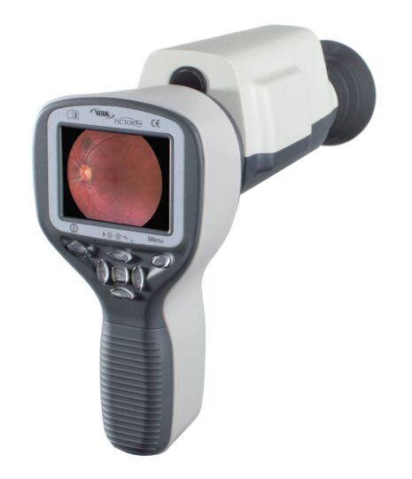
The Volk Pictor Plus™ portable ophthalmic camera can take your practice places. From the exam room to on-location screenings, nursing home calls and everywhere in between.
Two easily interchangeable modules provide high resolution retinal (non-mydriatic) or external eye imaging.
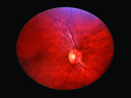
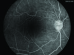
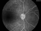
- Retinal Module – Pictor Plus retinal imaging enables non-mydriatic fundus examination with a 40º field of view. With digital still and video images, the appearance of optic disc, macula and retinal vasculature can be screened and documented for ocular lesions and anomalies.
- Anterior Module – Pictor Plus anterior imaging provides high-resolution images of the surface of the eye and areas directly surrounding the eye. The cobalt blue LED light allows fluorescent imaging to detect a dry eye or any trauma on the ocular surface.
Primary Functions
• Set up and import worklists into the camera
• Compare up to 9 simultaneous images displayed in the software
• Supported operating systems: Windows 7, Windows 8, & Windows 10
Key Benefits
• Wirelessly automate image transfer from Pictor Plus via Wi-Fi or USB
• Seamless workflow from imaging to diagnosis
• Operate as a standalone system for efficient data viewing, analysing and archiving
• Take advantage of complete tools for diagnostic purposes including zoom, lens and colour filtering
Interchangeable Modules
Two easily interchangeable modules provide high resolution retinal (non-mydriatic) or external eye imaging
Retinal Module
Pictor Plus retinal imaging enables non-mydriatic fundus examination with a 40° field of view. With digital still and video images, the appearance of optic disc, macula and retinal vasculature can be screened and documented for ocular lesions and anomalies.
- 9 fixation points to target a variety of retinal images
- Reflection‑free imaging with ten illumination levels
- Not necessary to dilate pupils (min. pupil size 3 mm)
- Produces colour and red‑free images for contrast
- Automatic and manual focusing
- Diopter compensation -20D to + 20D
- Slit lamp mount adapts Pictor to any slit lamp
Anterior Module
Pictor Plus anterior imaging provides high resolution digital image data of the surface of the eye and areas directly surrounding the eye. The cobalt blue LED light allows fluorescent imaging to detect a dry eye or any cuts or rashes on the surface of the eye.
- White and blue LED light for image targeting and capture
- Autofocus and optional manual adjustment
- 6x digital zoom
- Image resolution 2560 x 1920 pixels
To see our full range of Volk lenses, click here


