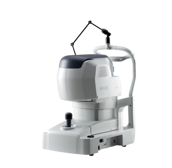
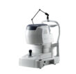
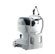
NIDEK RS-1 Glauvas OCT – Faster workflow without compromising diagnostic confidence
The NIDEK RS-1 Glauvas is an innovative OCT system with 250kHz scan speed, high-quality wide and deep area imaging, great operability, and deep learning-based analytics. With these capabilities, the RS-1 Glauvas provides streamlined workflow and diagnostic confidence for glaucoma and retinal vascular diseases in high-volume clinical practices.
250,000 A-scans/s high-speed imaging
The incorporation of 250,000 A-scans/s accelerates your workflow by reducing capture time. The high-speed imaging also addresses patient fixation errors thus contributing to greater image clarity and patient comfort.
Wide, deep, high resolution imaging
With the NIDEK RS-1 Glauvas, a single B-scan image clearly presents the area from the optic nerve head to the temporal vascular arcade, and the 4.2 mm depth B-scan imaging readily captures the oblate retinal shape of myopic eyes. Improvements in AngioScan OCT-Angiography include wider and clearer images for assessing chorioretinal microvasculature.
Effortless operation and interpretation
Easy image capture with automated functions
The auto alignment and auto switch functions allow anyone to effortlessly capture images. Operators need to only adjust the chinrest height and click Optimize and Capture.
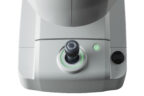
Newly-designed joystick for smooth operation
Based on user preference, operation with a newly-designed joystick is also available. The 4-direction button allows smooth, fine movement of the device.
Combo image capture for better clinic efficiency
Customizable combo image capture provides preset scanning patterns based on each target disease or examination routine of a facility, enhancing workflow.
Enhanced SLO sensitivity
The high SLO sensitivity facilitates reliable image capture, including in eyes with opacities, enabling preoperative assessment for cataract and diabetic retinopathy (DR).
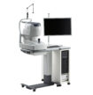
Intuitive user interface
The OCT Viewer software allows quick access to images and analytics. The display can be viewed by day, scan pattern, and analysis mode within a single screen – allowing faster review of imaging and analytics.
Structural Normality Map (SN Map) facilitating rapid interpretation
The SN Map detects and highlights structural abnormalities and even subtle retinal changes. This function clearly indicates a region of interest, layer by layer, and enhances clinical efficiency by reducing interpretation time.
Advanced Analytics
Glaucoma analysis in myopia
The long axial length normative database*1 presents analysis with axial length compensation, allowing for a more accurate glaucoma assessment in patients with axial myopia. The OCT Viewer automatically switches to this database as required, by using the axial length*2 which is a parameter for scan width correction.
Less false positives with deep learning segmentation (DL segmentation)
The accuracy of segmentation affects the outcomes of glaucoma analysis. DL segmentation reduces artifacts and errors in the normative database and thickness maps even in eyes with opacities, thus decreasing false positives and enhancing clinic efficiency by reducing unnecessary follow-up visits. Additionally, the scan width correction allows precise analytics based on the patient’s axial length*2.
*1 Data was collected from a sample of Asian patients.
*2 The value of the axial length is obtained based on the results of the OCT image capture and differs from the actual measured value of the axial length.
For full advanced analytics please refer to the product brochure