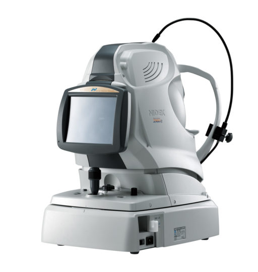
NIDEK Retina Scan Duo 2 OCT – Fast Combined HD OCT & Colour Fundus with NEW Denoising Software, Retina Map Scan & Optional OCT A.
High speed image capture
OCT images are captured at scan speeds of 70,000 A-scans/s which is 32% faster than acquisition with the Retina Scan Duo™ using Regular OCT sensitivity*
*Regular OCT sensitivity is used to capture images at high speed, and Ultra fine and Fine OCT sensitivity can be used to capture high definition images.
Wide area scan (12 x 9 mm)
Wide area normative database (macula: 9 x 9 mm, disc: 6 x 6 mm)
A 12 x 9 mm wide area image can be acquired. The retina map captures both the macula and disc in a single shot. The normative database provides a wide area color-coded map comparing the patient’s macular thickness to a population of normal eyes.
Denoising using deep learning
A new image enhancement technique using deep learning automatically displays a denoised image once B-scan acquisition is complete. With deep learning of a large data set of images averaged from 120 images, this denoising technique provides high definition images comparable to a multiple-image-averaging technique. The denoising function generates high definition images from a single frame while decreasing image acquisition time and increasing patient comfort.
Enhanced image
The image enhancement function allows adjustment of image brightness for advanced image quality and details.
Multiple OCT scan patterns
A wide range of scanning patterns allows selection of scans that suit the retinal region and ocular pathology.
Fundus Camera
12-megapixel CCD camera
The NIDEK Retina Scan Duo 2 includes a built-in 12-megapixel CCD camera, producing high quality fundus images with a 45° angle of view.
Stereo and panorama photography
The Retina Scan Duo™ 2 navigates stereo and panorama photography with target marks displayed on an observation screen, which enables the operator to easily capture stereo images and a panorama composition.
Fundus autofluorescence (FAF) *2
The FAF function is an advanced screening feature that allows non-invasive evaluation of the RPE without contrast dye. FAF is naturally emitted due to the presence of a substance called lipofuscin in the RPE cells. When stimulated with a specific wavelength of light, lipofuscin fluoresces and its distribution can be mapped.


